Retina
Retina is the innermost, thin layer of nerve cells that sends image to the brain for processing. The retinal diseases are some of the most common causes of a permanent loss of vision in patients, if not timely treated.


Common Retinal Diseases
1. Diabetic Retinopathy
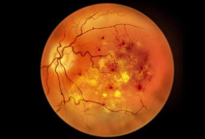
It is the most common disease of the retina, which damage the blood vessels of the retina and causes blindness.
Symptoms of Diabetic Retinopathy:
- Blurred vision
- Distorted vision
- Fluctuating vision
- Floaters
- Colour blindness
- Poor night vision
- Loss of vision
Types of Diabetic Retinopathy:
- Mild nonproliferative diabetic retinopathy.
- Moderate nonproliferative diabetic retinopathy.
- Severe nonproliferative diabetic retinopathy.
- Proliferative diabetic retinopathy.
- Mild nonproliferative diabetic retinopathy: Tiny blood vessels of the retina swell and bulge out. These are called microaneurysms.
- Moderate nonproliferative diabetic retinopathy: Tiny blood vessels further the swell and blocking blood flow to the retina. Blood and other fluids accumulate at the macula.
- Severe nonproliferative diabetic retinopathy: In this stage larger sections of blood vessels of retina become blocked. This lack of blood triggers a signal of the body to start growing new blood vessels in the retina. These new blood vessels are extremely thin and fragile and cause retinal swelling, blurry vision, dark spots and even patches of vision loss. If these vessels leak in to the macula sudden and permanent vision loss may occur.
- Proliferative diabetic retinopathy: At this advanced stage of the disease, new blood vessels continue to grow in the retina. Thin and week blood vessels are prone to bleeding and causes retinal scarring which eventually causes retinal detachment, which causes permanent blindness.
Treatment of Diabetic Retinopathy:
- Strict metabolic control
- Eye injections: Intra vitreal Anti-VEGF injections and steroid injections are given.
- Laser surgery: This procedure is called Panretinal photocoagulation which helps to shrink the abnormal blood vessels by giving laser burns.
- Vitrectomy: In advanced stages, vitrectomy is done remove blood, scar tissue, and some of vitreous gel. Retinal detachment can be corrected in the same way.
2. Retinopathy of Prematurity (ROP)
ROP is mainly caused due to retinal blood vessels developing abnormally and affects prematurely born babies. The abnormal proliferation of retinal blood vessels developed due to relative lack of oxygen.
Stages of ROP:
Stage 1: Demaracation line
Stage 2: Ridge
Stage 3: Ridge with the extraretinal fibrovascular proliferation
Stage 4: Retinal detachment “plus” disease (+)
Symptoms:
- Vision loss
- Visual field defects, usually in central vision
Risk factors:
New born babies with birth weight less than 1250 grams and gestational age less than 28 weeks.
ROP is commonly seen in low birth weight babies who received oxygen therapy.
Treatment:
- Intra vitreal Anti VEGF injections
- Photocoagulation with laser energy
3. Age related macular degeneration (ARMD)
ARMD is a common eye disease mostly seen in the people over the age of 50 years. This disease gradually destroys sharp, central vision.
Types:
- Dry ARMD: It is the most common type of ARMD in which there is slow atrophy of macula, leading to a gradual loss of central vision.
- Wet ARMD: It is less common but more severe disease than dry ARMD.
Risk factors of ARMD:
- Age: Older people are at more risk.
- Gender: Women are more affected then men.
- Genetics: Genetic predisposition is known in ARMD.
- Smoking: A definite risk factor.
- Sunlight: Over exposure to UV rays increases the risk of ARMD. Do wear sunglasses.
- Nutrition: Nutritious diet with supplementation with antioxidants and vitamins help protect again ARMD.
Diagnosis:
ARMD is detected by doing Optical coherence tomography (OCT)
Fluorescein angiogram
Treatment:
- Antioxidants
- Intra vitreal Anti VEGF injection
- Laser photocoagulation
- Photodynamic therapy
- Transpupillary thermotherapy
4. Retinal detachment (RD)
RD occurs when the retina is pulled away from its normal position. RD can cause permanent loss of vision if not treated in time.
Types:
- Rhegmatogenoud Retinal detachment: It occurs due to a hole or breaks in the retina
- Exudative retinal detachment: It occurs due to inflammation that results in fluid accumulating underneath the retina.
- Tractional retinal detachment: It occurs when fibrovascular tissue on the surface pulls the retina from the underlying tissues.
Symptoms:
- Flashes of light
- Floaters
- Shadow in the field of vision
- Loss of vision when macula is detached
Treatment:
- Scleral buckling: A silicone band is stitched around the eyeball to gently push the ball of the eye against detached retina.
- Vitrectomy: It is done to take out the vitreous gel , and is replaced with silicone oil or gases to push the retina in the place. Laser is around the breaks to seal it.
5. Macular hole
Macular hole is a small gap that opens at the centre of the retina which is called the macula. It could be present one eye or both eyes.
Symptoms:
- Blurred and distorted vision.
- Small blind spots in the centre of the vision.
- Total loss of vision.
Risk factor:
- Women at the age of 60 to 80 years.
- Vitreomacular traction.
- Retinal detachment.
- Eye injury.
- Swelling of the central retina (cystoid macular oedema).
Diagnosis:
Optical coherence tomography (OCT)
Treatment:
- Vitrectomy: Macular hole can be repaired using by operation called vitrectomy. Pars plana vitrectomy with tamponade done with or without peeling of internal limiting membrane. ILM flaps are used to fill the larger holes.

